A Collagen Patch May Help Dogs With a Stubborn Dental Problem
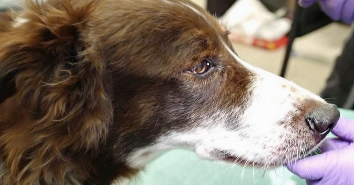
Dog oronasal fistula — a small hole that opens between a dog’s mouth and nasal cavity — is a painful and tricky condition to fix. But a new case report published in Frontiers in Veterinary Science offers some encouraging news. Six dogs with difficult-to-repair fistulas were treated using a collagen membrane (a thin, spongy patch made of protein) placed alongside standard surgery. All six dogs stopped sneezing and had their nasal discharge clear up within 14 to 21 days. None showed signs of the fistula coming back during follow-up of up to seven months.
While this is a very small study and cannot prove the membrane alone made the difference, the results add to what vets know about managing one of dentistry’s more challenging repairs.
What Is an Oronasal Fistula — and Why Does It Matter?
Picture a tiny tunnel running from the roof of your dog’s mouth straight into the bottom of their nose. That is essentially what an oronasal fistula is — an abnormal opening that connects the mouth and the nasal cavity above it. Food, water, and bacteria can pass through this opening and into the nose, causing constant irritation.
Dogs with this condition often show signs that look like a persistent cold:
- Sneezing that won’t go away
- Runny nose or nasal discharge, sometimes with traces of blood
- Recurring nasal infections
The most common cause is periodontal disease — the same tooth-and-gum disease that affects many dogs, especially older ones. When gum disease destroys the tissue and bone holding a tooth in place, a hole can form between the empty socket and the nasal passage above. The upper canine tooth — the large, fang-like tooth at the front of the mouth — is most often involved.
Surgery to close this opening is the standard fix, but it is notoriously tricky. The tissue in that area is tight, making a clean, secure closure hard to achieve. If the repair does not hold, the hole comes back.
Why Surgeons Are Exploring New Tools for This Repair
Standard oronasal fistula repair involves creating a tissue flap — a bit like cutting and sliding a small piece of gum tissue over the opening to seal it shut. Done well, this works. But in difficult cases — where the fistula is large, where previous repairs have failed, or where the surrounding tissue is thin or scarred — surgeons need extra support.
Collagen membranes are thin, spongy sheets made from collagen, a protein the body already produces naturally. In human dental surgery, these membranes are used as temporary scaffolds: they give healing tissue something to grow onto and protect the repair site while the body rebuilds. This study explored whether adding one of these membranes could improve outcomes in dogs with especially challenging fistulas.
How the Study Was Conducted
Researchers looked back at the records of six dogs — all client-owned pets seen at a veterinary dental or surgical practice — that had been treated for oronasal fistulas at the site of the upper canine tooth. Between the six dogs, there were eight fistula sites in total (some dogs had more than one opening to close).
Each dog had their fistula repaired using a single-layer tissue flap — gum tissue sutured over the hole — combined with a collagen membrane placed underneath to support healing from below. This approach is sometimes called guided tissue regeneration, a method borrowed from human dentistry to help direct how new tissue grows back.
Key details at a glance:
- Patients: 6 dogs, 8 fistula sites
- Location: All fistulas involved the upper canine tooth socket area
- Procedure: Tissue flap closure plus collagen membrane support
- Follow-up period: 21 days to 7 months after surgery
What the Results Showed
The outcomes were encouraging across all six dogs:
- Sneezing, nasal discharge, and nosebleeds resolved within 14 to 21 days after surgery
- No dog showed signs of the fistula returning at any follow-up visit
Whether follow-up lasted just three weeks or as long as seven months, none of the dogs had their symptoms come back. That is meaningful because recurrence — the hole reopening — is one of the biggest challenges with this repair. A technique that supports durable closure, even in tough cases, is worth noting.
The researchers noted, however, that sound surgical technique — especially careful flap design and making sure there is no tension pulling the repaired tissue apart — is just as important as the membrane itself. The collagen membrane is a support tool, not a substitute for skilled surgery.
What This Means for Your Dog
Good News for Difficult Cases
If your dog has been diagnosed with an oronasal fistula — especially one that has failed to heal after a previous repair, or that involves a large or complex opening — this research suggests that adding a collagen membrane to the standard procedure may be worth discussing with your vet. Veterinary dental specialists and oral surgeons are often familiar with these membranes because similar techniques are used in other parts of veterinary and human dentistry.
The Best Protection Is Prevention
The most actionable takeaway for most dog owners is this: keep up with dental care. Oronasal fistulas most often develop because periodontal (gum and tooth) disease has gone unchecked for too long. Regular dental check-ups, professional cleanings, and home dental care can catch gum disease early and protect the bone that supports the teeth.
Watch for these signs and mention them to your vet:
- Bad breath that has gotten noticeably worse
- Dropping food or chewing on one side
- Pawing at the mouth or face
- Sneezing more than usual, or a runny nose that won’t clear up
When to Ask About a Specialist
If your vet suspects an oronasal fistula — or if a repair has already failed — it is reasonable to ask for a referral to a board-certified veterinary dentist or oral surgeon. These specialists perform this type of repair regularly and have access to techniques and materials like collagen membranes that can improve the odds of lasting success.
Limitations to Keep in Mind
This study is very small. Only six dogs were included, and it was a retrospective case series — meaning researchers reviewed existing records rather than running a planned experiment with a comparison group. There was no group of dogs treated without the membrane, so it is not possible to say for certain that the membrane itself drove the good outcomes. Good surgical skill — particularly careful flap design and tension release — is just as critical, as the researchers themselves acknowledged.
Follow-up times also varied widely, from just three weeks for some dogs to seven months for others. A longer, more standardized follow-up across all cases would give a clearer picture of true long-term success rates.
Larger, controlled studies are needed before this approach can be considered standard practice.
The Bottom Line
For dogs with difficult oronasal fistulas, adding a collagen membrane to standard tissue flap surgery looks promising. In this small case series, all six dogs cleared up their symptoms within three weeks, and none showed recurrence during follow-up of up to seven months. The collagen membrane acts as a scaffold to support healing, but it works alongside — not instead of — skilled surgical technique.
For most dog owners, the clearest action step is routine dental care. Catching and treating gum disease early is the best way to prevent the tooth and bone loss that leads to this condition in the first place.
This article summarizes peer-reviewed research for educational purposes. Always consult with your veterinarian for personalized advice about your pet’s health and behavior.